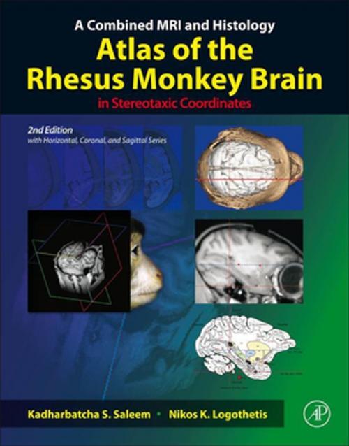
A Combined MRI and Histology Atlas of the Rhesus Monkey Brain in Stereotaxic Coordinates
Nonfiction, Health & Well Being, Medical, Specialties, Internal Medicine, Neurology, Science & Nature, Science, Biological Sciences| Author: | Kadharbatcha S. Saleem, Nikos K. Logothetis | ISBN: | 9780123850881 |
| Publisher: | Elsevier Science | Publication: | September 1, 2012 |
| Imprint: | Academic Press | Language: | English |
| Author: | Kadharbatcha S. Saleem, Nikos K. Logothetis |
| ISBN: | 9780123850881 |
| Publisher: | Elsevier Science |
| Publication: | September 1, 2012 |
| Imprint: | Academic Press |
| Language: | English |
A Combined MRI and Histology Atlas of the Rhesus Monkey Brain in Stereotaxic Coordinates, Second Edition maps the detailed architectonic subdivisions of the cortical and subcortical areas in the macaque monkey brain using high-resolution magnetic resonance (MR) images and the corresponding histology sections in the same animal. This edition of the atlas is unlike anything else available as it includes the detailed cyto- and chemoarchitectonic delineations of the brain areas in all three planes of sections (horizontal, coronal, and sagittal) that are derived from the same animal. This is a significant progress because in functional imaging studies, such as fMRI, both the horizontal and sagittal planes of sections are often the preferred planes given that multiple functionally active regions can be visualized simultaneously in a single horizontal or sagittal section. This combined MRI and histology atlas is designed to provide an easy-to-use reference for anatomical and physiological studies in macaque monkeys, and in functional-imaging studies in human and non-human primates using fMRI and PET.
- The first rhesus monkey brain atlas with horizontal, coronal, and sagittal planes of sections, derived from the same animal
- Shows the first detailed delineations of the cortical and subcortical areas in horizontal, coronal, and sagittal plane of sections in the same animal using different staining methods
- Horizonal series illustrates the dorsoventral extent of the left hemisphere in 47 horizontal MRI and photomicrographic sections matched with 47 detailed diagrams (Chapter 3)
- Coronal series presents the full rostrocaudal extent of the right hemisphere in 76 coronal MRI and photomicrographic sections, with 76 corresponding drawings (Chapter 4)
- Sagittal series shows the complete mediolateral extent of the left hemisphere in 30 sagittal MRI sections, with 30 corresponding drawings (Chapter 5). The sagittal series also illustrates the location of different fiber tracts in the white matter
- Individual variability - provides selected cortical and subcortical areas in three-dimensional MRI (horizontal, coronal, and sagittal MRI planes). For comparison, it also provides similar areas in coronal MRI section in six other monkeys. (Chapter 6)
- Vasculature - indicates the corresponding location of all major blood vessels in horizontal, coronal, and sagittal series of sections
- Provides updated information on the cortical and subcortical areas, such as architectonic areas and nomenclature, with references, in chapter 2
- Provides the sterotaxic grid derived from the in-vivo MR image


