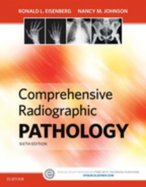
Comprehensive Radiographic Pathology - E-Book
Nonfiction, Health & Well Being, Medical, Allied Health Services, Radiological & Ultrasound| Author: | Ronald L. Eisenberg, MD, JD, FACR, Nancy M. Johnson, MEd, RT(R)(CV)(CT)(QM), FASRT | ISBN: | 9780323370257 |
| Publisher: | Elsevier Health Sciences | Publication: | July 29, 2015 |
| Imprint: | Mosby | Language: | English |
| Author: | Ronald L. Eisenberg, MD, JD, FACR, Nancy M. Johnson, MEd, RT(R)(CV)(CT)(QM), FASRT |
| ISBN: | 9780323370257 |
| Publisher: | Elsevier Health Sciences |
| Publication: | July 29, 2015 |
| Imprint: | Mosby |
| Language: | English |
Gain the essential pathology understanding you need to produce quality radiographic images! Covering the disease processes most frequently diagnosed with medical imaging, Comprehensive Radiographic Pathology, 6th Edition is the perfect pathology resource for acquiring a better understanding of the clinical manifestation of different disease processes, their radiographic appearances, and their treatments. This full-color reference begins with a general overview of physiology, then covers disorders and injuries by body system. The new edition also includes the latest information on CT, MRI, SPECT, PET, ultrasound, and nuclear medicine — including updated radiographer notes, images, and review questions.
-
Thorough explanations and comprehensive coverage aid readers’ understanding of disease processes and their radiographic appearance.
-
Numerous high-quality illustrations covering all modalities clearly demonstrate the clinical manifestations of different disease processes and provide readers with a standard for the high-quality images needed in radiography practice.
-
Discussion of specialized imaging explains how supplemental modalities, such as ultrasound, computed tomography, magnetic resonance imaging, nuclear medicine, single-photon emission computed tomography (SPECT), and positron emission tomography (PET) are sometimes needed to diagnose various pathologies.
-
Treatment coverage provides readers with brief explanations of the most likely treatments and the prognosis for each pathology.
-
Systems-based approach organizes the pathology of various body systems in separate chapters — each chapter provides an initial discussion of general physiology and then explains various pathologic conditions and their radiographic appearance and treatment.
-
Summary Findings tables are a great quick reference guide for practitioners.
-
Consistent organization aids readers in searching for information.
-
Study aids include an outline, key terms, objectives, and review questions for every chapter.
-
Useful appendices include an extensive glossary; a list of major prefixes, roots, and suffixes with definitions and examples; and a table of diagnostic implications of abnormal lab values.
-
NEW! Updated images in all modalities keep readers abreast on the latest advances needed for clinical success.
-
NEW! Updated chapter review questions have been added to the end of every chapter.
-
NEW! Additional review questions on Evolve companion site provide students with extra resources to prepare for certification.
-
**NEW! Updated radiographer notes **incorporate current digital imaging information for both computed radiography and direct digital capture.
