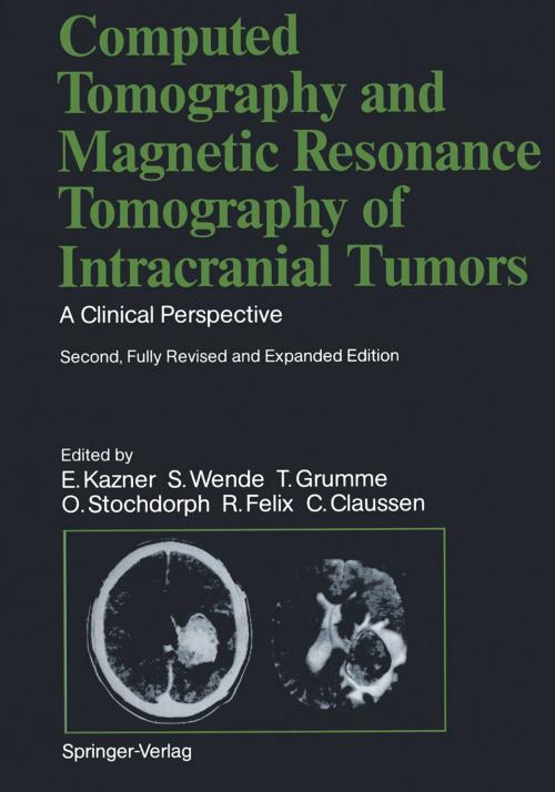
Computed Tomography and Magnetic Resonance Tomography of Intracranial Tumors
A Clinical Perspective
Nonfiction, Health & Well Being, Medical, Surgery, Neurosurgery, Specialties, Internal Medicine, Neurology| Author: | C. Claussen, R. Fahlbusch, R. Felix, T. Grumme, J. Heinzerling, J.R. Iglesias-Rozas, E. Kazner, K. Kretzschmar, M. Laniado, W. Müller-Forell, T.H. Newton, W. Schörner, G. Schroth, B. Schulz, O. Stochdorph, G. Sze, S. Wende, W. Lanksch | ISBN: | 9783642743115 |
| Publisher: | Springer Berlin Heidelberg | Publication: | December 6, 2012 |
| Imprint: | Springer | Language: | English |
| Author: | C. Claussen, R. Fahlbusch, R. Felix, T. Grumme, J. Heinzerling, J.R. Iglesias-Rozas, E. Kazner, K. Kretzschmar, M. Laniado, W. Müller-Forell, T.H. Newton, W. Schörner, G. Schroth, B. Schulz, O. Stochdorph, G. Sze, S. Wende, W. Lanksch |
| ISBN: | 9783642743115 |
| Publisher: | Springer Berlin Heidelberg |
| Publication: | December 6, 2012 |
| Imprint: | Springer |
| Language: | English |
This book represents the second, fully revised edition of the original volume published in 1982. Experience in neuroradiology has confirmed the outstanding value of computed tomography (CT) for the diagnosis of space-occupying lesions within the skull and orbit. It might be assumed, then, that the second edition of this book would simply represent a numerically expanded continua tion of the popular first edition. That is not the case, however. Advances in imaging techniques have promp ted the creation of a new book whose expanded title reflects its more comprehen sive nature. The added illustrations, the revised text, and the expanded circle of editors and contributors document this. Since publication of the first edition, a new modality, magnetic resonance imaging (MRI), has become an established neuroradiologic study. We felt it was essential to include this new modality in our book and explore its capabilities as an adjunct or alternative to CT scanning. Because of the high acquisition costs of MRI and the still small number of MR units currently in operation, we have relied in part on images furnished by other institutions and private practitioners, to whom we are indebted. Many problems relating to MR, both in terms of equipment and image interpretation, have yet to be resolved. There is no denying that we still have much to learn.
This book represents the second, fully revised edition of the original volume published in 1982. Experience in neuroradiology has confirmed the outstanding value of computed tomography (CT) for the diagnosis of space-occupying lesions within the skull and orbit. It might be assumed, then, that the second edition of this book would simply represent a numerically expanded continua tion of the popular first edition. That is not the case, however. Advances in imaging techniques have promp ted the creation of a new book whose expanded title reflects its more comprehen sive nature. The added illustrations, the revised text, and the expanded circle of editors and contributors document this. Since publication of the first edition, a new modality, magnetic resonance imaging (MRI), has become an established neuroradiologic study. We felt it was essential to include this new modality in our book and explore its capabilities as an adjunct or alternative to CT scanning. Because of the high acquisition costs of MRI and the still small number of MR units currently in operation, we have relied in part on images furnished by other institutions and private practitioners, to whom we are indebted. Many problems relating to MR, both in terms of equipment and image interpretation, have yet to be resolved. There is no denying that we still have much to learn.


