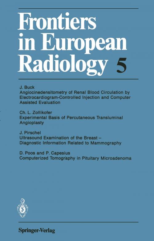
Frontiers in European Radiology
Nonfiction, Health & Well Being, Medical, Medical Science, Biochemistry, Allied Health Services, Radiological & Ultrasound| Author: | J. Buck, C.L. Zollikofer, J. Pirschel, D. Poos, P. Capesius | ISBN: | 9783642725630 |
| Publisher: | Springer Berlin Heidelberg | Publication: | December 6, 2012 |
| Imprint: | Springer | Language: | English |
| Author: | J. Buck, C.L. Zollikofer, J. Pirschel, D. Poos, P. Capesius |
| ISBN: | 9783642725630 |
| Publisher: | Springer Berlin Heidelberg |
| Publication: | December 6, 2012 |
| Imprint: | Springer |
| Language: | English |
Considerable advances have been made over the years in the study of the physiological and diseased states of the kidney, so that our present-day diagnostic capabilities permit not only morphological, but also functional inter-relations to be registeres. The first step toward function diagnostics was taken with the introduction of kymography. This was followd by serial angiography, and then came cineradiography which made simultaneous morphological and functional radiological examinations feasible. 2 Physiology of Kidney 2. 1 Hemodynamics of Renal Arteries Because of the negligible vascular elasticity of the rani arteries, the diameter variation resulting within one cardiac action is about 1 % [14]; but with these variations being lost in the accuracy of measurement, however, blood flow can be compared to the flow of liquid in rigid tubes. In a rigid tube, the velocity of the individual particles of liquid varies in relation to its distance from the tube axis. Under normal circumstances, a laminar flow prevails in the arteries, the blood flowing parallel to the vascular wall in coaxial cylindrical layers. The velocity is referred to here as v. The flow rate, I, stands for the ratio of the volume of liquid, V, flowing through a cross section of a tube to the time, t, required for this, or also for the product of flow velocity v and the cross section of the tube q: 1= Llv: Llt and I = v x q.
Considerable advances have been made over the years in the study of the physiological and diseased states of the kidney, so that our present-day diagnostic capabilities permit not only morphological, but also functional inter-relations to be registeres. The first step toward function diagnostics was taken with the introduction of kymography. This was followd by serial angiography, and then came cineradiography which made simultaneous morphological and functional radiological examinations feasible. 2 Physiology of Kidney 2. 1 Hemodynamics of Renal Arteries Because of the negligible vascular elasticity of the rani arteries, the diameter variation resulting within one cardiac action is about 1 % [14]; but with these variations being lost in the accuracy of measurement, however, blood flow can be compared to the flow of liquid in rigid tubes. In a rigid tube, the velocity of the individual particles of liquid varies in relation to its distance from the tube axis. Under normal circumstances, a laminar flow prevails in the arteries, the blood flowing parallel to the vascular wall in coaxial cylindrical layers. The velocity is referred to here as v. The flow rate, I, stands for the ratio of the volume of liquid, V, flowing through a cross section of a tube to the time, t, required for this, or also for the product of flow velocity v and the cross section of the tube q: 1= Llv: Llt and I = v x q.


